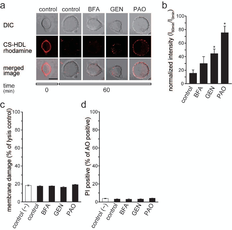
Fig. 7. Cell membrane localization of TC1-cpHDL at 60 min after photo-induced depolarization. (a) DIC (differential interference contrast) and confocal microscopy images of PC12 cells treated with TC1-cpHDL rhodamine (CS-HDL rhodamine), and treated with BFA, GEN, and PAO at 0 min (left panel) and 60 min. Filled bar indicates 10 µm. (b) The normalized intensities of rhodamine-positive lines of 10 total lines (see Materials and Methods) at 0 min and 60 min after 2-min illumination (hν: 525−550 nm, input power 2 mW cm−2) are shown for PC12 cells treated with BFA, GEN, and PAO. *p<0.05 vs. control (n = 5). (c) Lactate dehydrogenase (LDH) assay for PC12 cells without TC1-cpHDL (control) and with TC1-cpHDL (black bar) treated with BFA, GEN, and PAO at 60 min after 2-min illumination (525−550 nm, input power 2 mW cm−2). (d) Acridine Orange/Propidium Iodide (AO/PI) assay in PC12 cells without TC1-cpHDL (white bar) and with TC1-cpHDL (black bar) treated with BFA, GEN, and PAO at 60 min after 2-min illumination (525−550 nm, input power 2 mW cm−2).